Cell Biology: Mitochondria | General
Cell Membrane
Phospholipid Bilayer.
Dynamic fluid model
FRAP: Fluorescence Recovery After Photobleaching
 |
|---|
| © Niklas Lorén, et al. |
Prokaryotes and Eukaryotes
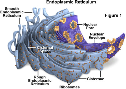
Cells that have internal membranes are called Eukaryotes.
 |
|---|
| © Michael W. Davidson and The Florida State University |
The layer of the ER is Cisternae. The space between the Cisterna is the lumen.
Mitochondria
Cardiolipin is an abundant lipid in the inner membrane but not in the outer membrane.
The inner membrane is highly impermeable to small ions.
Three classes of protein that makeup 76% of mass:
- critical for catalyzing a subset of reactions necessary for metabolism.
- transport protein
- F1F0APTase
Matrix: Enzyme for the citric cycle.
Moving of the Mitochondria
That microtubule ware moves along the microtubule network. Those microtubules were remodeling and dynamically to respond the metabolic change or metabolic stress. As a result, the mitochondria can relocate through this work.
The distribution of the mitochondria is diverse and depends on the type of the cell. For example, in neurons, mitochondria are rich in Axon Hillock[1] areas.
Endosymbiosis: living together.
Mitochondria Disease
A cell could have a few or a more different set of mitochondria. As a result, people may look healthy though they carry malfunction mitochondria.
Sometimes, the carrier who has enough healthy mitochondria could still cause problems. For example, her infant could be fetal because, during the egg division, one egg may randomly receive more malfunction-mitochondria and leading mitochondria diseases.
Observed Evidence: (Threshold of mitochondria mutation)
- 20~40% of malfunction: you can still generate enough ATP.
- 50~60% of malfunction: disease presentation.
3-way IVF (3-way in vitro fertilization)
Fertilization in a dish.
- Extract the nucleus from the mother and inject it into a donor egg.
In this way, you’ll have three biology parents:
sperm from your father. nucleus gene from your mother; mitochondria gene from donor mother. (It was approved in the UK but not in the US)
Electronic Transport Chain
Why electronic transport. The inner membrane is low-permeable for protons which could help to sustain a high proton gradient. The proton is the key to driving the F1F0APTase to synthesis the ATP by pumping the proton out of the matrix. Without the proton gradient, the ATP could not be generated and the cell would die.
General:
- Complex I, III, and IV: directly pump the proton into the intermembrane space (have both electron carrier and proton pump).
- Complex II can promote the activity of complex III and IV. (has electron carrier only)
They get the energy for pumping by transferring electrons through a series of coupled reactions. This linked process is what we call the electron transport chain.
- Complex I:
NADH deposit two high-energy electrons and passed along Redox Centers.
Redox Center:- The bottom of the center has high affinity than the top.
- The adjacent redox center is ideal for electron jump.
- small energy was released by each jump which would be used to pump protons.
- The last redox center was passed two electrons to a Coenzyme Q
- Complex II:
Similar to the complex I.- High energy was released from FADH2
- Electron was delivered into Coenzyme Q through the redox center.
- Difference: The liberated energy doesn’t use to pump protons.
- Complex III:
- The electrons were released from Coenzyme Q
- one electron was recyclable, the other will pass through two redox centers before reaching cytochrome C
- cytochrome C carry the electron into Complex IV
- Complex IV:
- four electrons convert a molecule of oxygen to two molecules of water.
Coenzyme Q is hydrophobic and diffusion inner the bilayer.
Cytochrome C is a peripheral protein and is associated with the inner face of the inner face membrane.
More details at Biochemistry: Electronic transport
Metabolism
Term:
Electron donor, Electron Acceptor. (O2 is the final electron accepter)
Oxidation, Reduction
3 Steps of metabolism
Electron transport
The proton pump transported protons from matrix to Intermembrane space which made the Matrix side is basic, the intermembrane side is acidic.
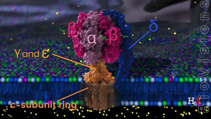
F1F0APTase
Binding Change Model
It is an F-class pump
 |
|---|
| © HavardX |
When the C-subunit ring is rotating, the $\gamma$ and $\epsilon$ unit are rotated with c-subunit. However, the $\alpha,\ \beta,$ and $\delta$ was not rotating.
$\delta$ subunit is bound to B subunit which is embedded into the inner mitochondria membrane.
Binding Change Model
-
Open state, Loss state, Tight state
Conformation switches with the rotating of the $\gamma$ subunit.
or initial segment. This is the region where the plasma membrane generates nerve impulses; the axon conducts these impulses away from the soma or dendrites toward other neurons. Britannica ↩︎
Cell Biology: Mitochondria | General
https://karobben.github.io/2021/08/17/LearnNotes/edx-cellbio-1/


