
2 Membranes|Advanced Cell Biology|Tulane
| Proof Reading: Haoyang Liang |
|---|
Overview
- A cell is a membrane-rich environment
- Classes of lipids
- Structure of lipids
- Polymorphic phase behavior of lipids
- The lipid bilayer membrane
- The fluid mosaic model of biological membranes
- Classes and functions of membrane proteins
- Receptors
- Hydropathy plots
Terminology
phospholipid bilayers:
- amphipathic: hydrophobic tial and hydrophilic head
- micelles; liposomes; sheet-like phospholipid bilayers
Function
- Selective Barrier
- Surround cells to hold enzymes and metabolites inside
- Cellular Compartmentation: surround organelles
- Contain Enzyme Systems–energy metabolism
(oxidative phosphorylation, photosynthesis, etc.)- Contain Transport Systems–bring food molecules inside and maintain ion concentrations
- Contain Specific Recognition Sites–for hormones, etc.
PS:
- Reaction Facility: Enzyme Working on a 3 dimension, -> with the membrane, bring things to 2 dimensions to facilities the interactions and reactions.
- protein helps to transport the materials.
- hydrophobic substrate; recognition: Glycolipids / Glucoportein
Composition
- Lipids – lipid bilayer creates a hydrophobic barrier mostly phospholipids but also glycolipids (contain carbohydrate) and steroids (cholesterol). Lipid bilayers form a barrier to the diffusion of hydrophilic molecules.
- Proteins – confer specificity
- peripheral membrane proteins bound to bilayer surface
- integral membrane proteins – intrinsic structural parts with hydrophobic and hydrophilic domains ===> amphipathic
- glycoproteins (integral) contain covalently bound CH2O which may be very complex ===> cell surface receptors?
e.g. glycophorin–MW 30,000 with 130 aa’s and 60% CH2O
- Exceptions: drugs which specifically designed to cross membrane to deliver the chemicals.,
- hydrophilic for solving
- hydroponically for crossing the membrane
The relative amounts of lipid and protein vary from one membrane type to the next, but the basic structures of all of these membranes are similar.
| Membrane | Lipid | Protein |
|---|---|---|
| Myelin Sheath | 80% | 20% |
| Plasma Membrane | 50% | 50% |
| Mitochondrial Inner Membrane | 25% | 75% |
The ratio of the L/P of the membrane serves its function.
Structural of lipid
Phospholipids
Hydrophilic head
Hydrophobic tail
Saturated tail/Unsaturated tail
Unsaturated tail:
- Decreases the Van de Waal interaction by increasing the distance of the lipid tail.
- increases fluidity.
Cholesterol
Abundant in mammal’s cell but lack in prokaryotic cells.
Maintain the fluidity and rigidity of the cell membrane[1]
Modulating the physical property of the membrane.
Large spacial molecular, decreasing fluid-ability increasing the order of the membrane
Lipid Bilayers
Free rotation and diffusion in 2-dimensions
Micelle
Vesicle
Classes
- Fatty acid (e.g. palmitate, oleate)
- Energy storage metabolite
- Building block of other lipids
- Triacylglycerols (fat)
- Energy storage
- Glycerophospholipids
- Form lipid bilayer membrane
- Signal transduction
- Sphingolipids
- Form lipid bilayer membranes
- Signal transduction
- Steroids
- Rigidify membranes (Cholesterol)
- Membrane-crossing hormones (e.g. Estrogen)
Non-pivotal:
classification:
- head
- Tail
“Fluid Mosaic” model of biological membranes
 |
|---|
| © Veronika Novotná, et al. |
“The membrane (is) a two-dimensional liquid-like solution of protein dissolved in the lipid bilayer”
Cite: Singer SJ, Nicolson GL The fluid mosaic model of the structure of cell membranes Science 1972; 175(23):720-3
Membrane Composition
Asymmetry of the membrane composition in phospholipid, protein, and sterol.
Integral Membrane Proteins
- α-spin in the interior of the bilayer,
- = =
two known structural motifs in membrane
- $\beta$-barrel
- Outer membrane
- Mitochondria membrane
E. Coli Outer Membrane Protein A (OmpA) an 8-stranded β-barrel. Known variation: 8-22 strands. About 2-4% of genome of Gram negative bacteria. Less than 1% of mitochondrial proteins
- Helical Bundle
- rich in eukaryotic cells.
Bacteriorhodopsin (mitochondrial), a 7-helix helical bundle protein. Known variation 1-22+ helices. About 25% of the proteins of ALL known genomes are helical membrane proteins.
Classes of Helical
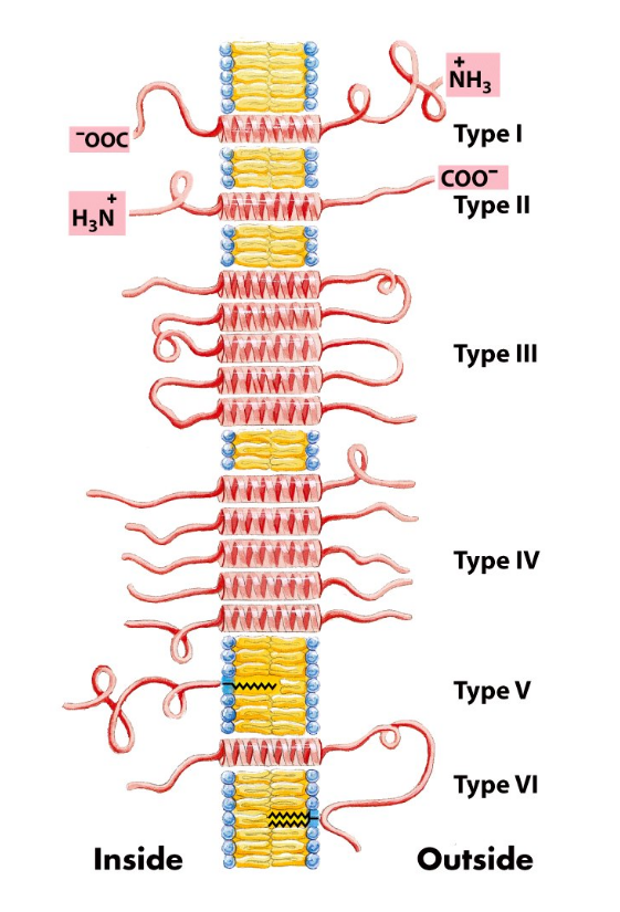
Type I and II -> opposite in direction
- I: Out -> inner (N -> C)
- II: Out -> inner (C -> N)
Type III: single back-forward (snake-like)
Type IV: a cluster of multiple bundles
Type V: no-helix with lipids
Type VI: helix + lipid(s) linkage
FRAP
FRAP - Fluorescence Recovery after Photobleaching
A technique used to measure protein diffusion in membranes. FRAP uses a fluorescence microscope to measure the diffusion of molecules into a photobleached spot in a membrane.
A membrane component can be freely diffusing, coralled or immobile
Functional Classes
Enzymes - catalyze reactions with lipid soluble substrates
Channels/pores - Allow molecules to move across membranes
Transporters - Pump molecules across membranes
Toxins - Permeabilize membranes to kill cells
Binding Proteins - Regulation, stabilization/destabilization, activation/deactivation, signal transduction
Anchors - Attachment point for cell-surface carbohydrates or for cytoskeletal proteins
Transporters
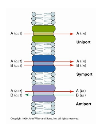
Some types of protein mediated transport across membranes.
Symporters and antiporters can pump one of the solutes against a concentration gradient
Some uniporters use metabolic energy to pump solutes against a concentration gradient. Others only allow the flow of solutes down a concentration gradient.
Receptors
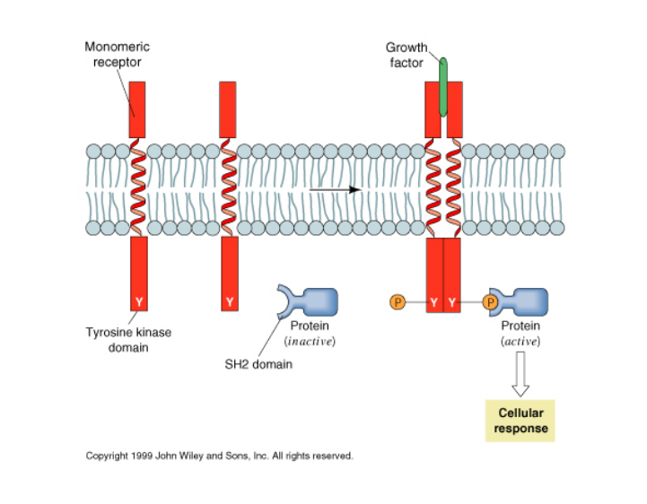
A membrane-spanning receptor transduces chemical signal across membranes
Some broad classes:
Channel receptors (Acetylcholine receptor)
Tyrosine Kinase (Insulin, growth factor)
G-protein coupled (adrenergic, rhodopsin)G-protein coupled receptors all belong to a widespread class called 7TM receptors because they have 7 membrane-spanning helices.
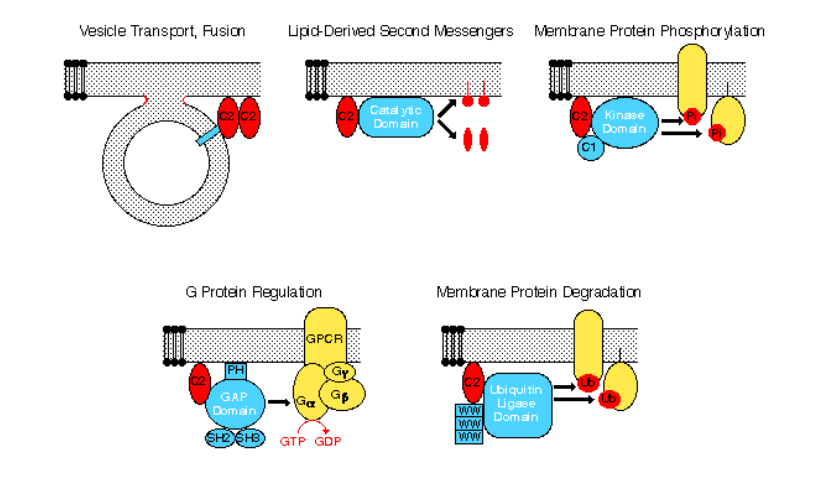
The C2 Domain
- sterols provide structural support to membranes, preventing too close a packing of the phospholipids’ acyl chains to maintain a significant measure of membrane fluidity, and at the same time conferring the necessary rigidity required for mechanical support. 2. Cholesterol restricts the random movement of phospholipid head groups at the outer surfaces of the leaflets, but its effect on the movement of long phospholipid tails depends on its concentration. At the cholesterol concentrations normally present in the plasma membrane, the interaction of the steroid ring with the long hydrophobic tails of phospholipids tends to immobilize those lipids and thus decreases biomembrane fluidity. It is this property that can help organize the plasma membrane into discrete subdomains of unique lipid and protein composition. At lower cholesterol concentrations, however, the steroid ring separates and disperses phospholipid tails, causing the inner regions of the membrane to become slightly more fluid. [Molecular Cell Biology, 8th, Harvey Lodish, et al.]
2 Membranes|Advanced Cell Biology|Tulane
https://karobben.github.io/2021/08/26/LearnNotes/tulane-cellbio-2/


