
5 Membrane Transport|Advanced Cell Biology|Tulane
Objectives
By the end of this session you should be able to:
- Compare and contrast the characteristics of
- passive diffusion
- facilitated diffusion
- primary active transport
- secondary active transport (cotransport).
- Compare and contrast the properties of the:
- four classes of ATP-powered pumps
- their effect on the intracellular environment
- Describe how:
- potassium channels can generate a hyperpolarized resting potential, common in most living cells.
- Explain how
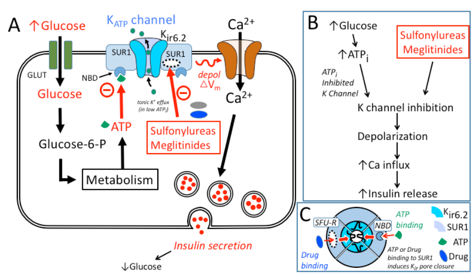
- transporters & Ion channels regulate insulin release in the pancreas
General information
Type of membrane transport depends on:
- Characteristics of the substance being transported
- Direction of transport
Three main classes of membrane protein
- ATP-power pump (carrier, permease) couple with energy source for active transport binding of specific solute to transporter which undergo conformation change.
- Channel protein (ion channel) formation of hydrophilic allow passive movement of small inorganic molecule.
- Transporters:
- uniport (single)
- Symport (double)
- antiport (double & anti one cis another trans gradient)
 |
|---|
| © Mariafrancesca Scalise, et al. |
Two types of transport:
 |
|---|
| © Maria Müller |
- Passive transport (diffusion): NO metabolic energy is needed
- Active Transport: Metabolic energy is used
Passive (simple) diffusion
- Small noncharged molecules or lipid soluble molecules pass between the phospholipids to enter or leave the cell
- Moving from areas of high concentration to areas of low concentration (they move down their concentration gradient)
- Example: Oxygen, carbon dioxide, and most lipids
Osmosis
A type of simple diffusion in which water molecules diffuse through a selectively permeable membrane from areas of high water concentration to areas of lower water concentration.
Facilitated diffusion
- The substance is moving down its concentration gradient through a membrane protein (not between the phospholipids)
- Example: monosaccharide, glucose, water
- “Water & oil don’t mix”
- That’s why water was not considered as passive diffusion
- Aquaporins increase water permeability
- Too much mRNA injected into Frog Oocyte could make it absorbing too much water.
 |
|---|
| © Rob Horsefield, et al. |
GLUT1 Mechanism (Facilitated Diffusion)
 |
|---|
| © Dong Deng, et al. |
- most commonly expressed glucose transporter
- reversible (depends on concentration gradient); glucose is commonly rapidly phosphorylated in cytoplasm
- ping pong mechanism (protein does not “flip”)
Facilitated vs. Passive Diffusion
 |
|---|
| © Florina Silvia Iliescu, et al. |
Exp: GLUT1 (erythrocytes) and GLUT2 (liver & pancreas)
- saturable
- non-linear
- selective, fast & constant rate at 5-10 mM
- uptake rate varies more (stimulates insulin release)
Active transport
PS: Energy; Against Concentration
- Move substances across membranes against their concentration gradient (from areas of low concentration to areas of high concentration)
- Energy is needed for this movement
Primary vs Secondary active transport
| © Alberto Canarini, et al. |
- Primary: simply against the concentration
- Secondary: send the intermediate molecule against the concentration and then, Symport the target molecules against the concentration.
e.g.:
- Intestinal & Kidney
Utilize the free-energy for Na (down its concentration gradient) to move glucose (against its concentration gradient)- SGLT-2 (Na-glucose co-transporter 2)
Comparison of Different Transport Mechanisms
| Property | Passive Diffusion |
Facilitate diffusion |
Active Transport |
cotransport |
|---|---|---|---|---|
| Protein Required | - | + | + | + |
| Against Graident | - | - | + | + |
| ATP Needed | - | - | + | - |
| contransported ion down its gradient | - | - | - | + |
| EXP | O2, CO2, steroid hormones, may drugs | Glucose and AA (uniporters), ions, water (channels) | Ions, small hydrophilic molecules, lipids (ATP-powered pumps) | Glucose and AA (symporters); Various ions and sucrose (antiporters) |
- Saturable transport at high [solute]
4 Classes of ATP-powered transport proteins
- P class pump
- V class pump
- F Class Proton Pumps
- ABC (ATP Binding Cassette) Superfamily
P Class pump
(Proton pump)
- Plasma membrance of plants, fungi, bacteria (H+ pump)
- Plasma membrane of higher eukaryotes (Na+/K+ pump)
- Apical plasma membrane of mammalian stomach (H+/K+ pump)
- Plasam membrane of all eukaryotic cells (Ca+ pump)
- Sarcoplasmic reticulum membrane in muscle cells (Ca+ pump)
Typical Intracellular and Extracellular Ion Concentrations
| Ion | Cell(mM) | Blood (mM) |
|---|---|---|
| K+ | 139 | 4 |
| Na+ | 12 | 145 |
| Cl- | 4 | 116 |
| HCO3- | 12 | 29 |
| X- | 138 | 9 |
| Mg2+ | 0.8 | 1.5 |
| Ca2+ | <0.0002 | 1.8 |
Na/K ATPase
 |
|---|
| © Susan Spiller, et al. |
Inhibited by ouabain &
digoxin
F-Class Proton Pumps
Location:
- Bacterial plasma membrane
- Inner mitochondrial membrane
- Thylakoid membrane of chloroplast
- Use H+ gradient to generate ATP (convert chemical energy to a different form)
V-Class Proton Pumps
Location:
- Vacuolar membranes in plants; yeats, other fungi
- Endosomal and lysosomal membranes in animal cells
- Plasma membrane of osteoclasts and some kidney tubule cells.
- Utilize ATP to generate a H+ gradient
PS: Functional against F-Class
V-Class H+ ATPase
Lysosomes:
- degrade extracellular materials (e.g. bacteria, LDL particles)
Synapse:
- V-class H+ pumps & Synaptic Transmission
- Vesicles, Pumps & Synaptic Transmission
ATP Binding Cassette (ABC) Superfamily
- Bacterial plasma membrane (AA, sugar, and peptide transporters)
- Mammalian plasma membranes (Transporters of phospholipids, small lipophilic drugs, cholesterol, other small molecules)
- Pre-systemic drug clearance, blood-brain barrier, renal secretion of drugs, cancer cells
Location:
BBB (blood-brain barrier ); GI tract; Kidney; Liver; Cancer
ABC Transporters & Presystemic Clearance
Mechanism of presystemic clearance. After drug enters the enterocyte, it can undergo metabolism, excretion into the intestinal lumen, or transport into the portal vein. Similarly, the hepatocyte may accomplish metabolism and biliary excretion prior to the entry of drug and metabolites to the systemic circulation.
Ion Channel
Four Subtypes of Ion Channels
| Types | Example | |
|---|---|---|
| 1 | Voltage-gate | Na, Ca, K |
| 2 | Ligand-gated (extracellular ligand) |
GABAA, ACh, Nicotinic |
| 3 | Ligand-gated (intracellular ligand) |
βγ, Muscarinic, Adenosine |
| 4 | Stress-activated | Stretch |
 |
|---|
| © Nilan Jacob |
Beta Cells of the Pancreas
(How Transporters & Ion Channels Regulate Insulin Release)
 |
|---|
| © Suttira “Joy” Intapad, Ph.D |
Ion Channels & Membrane Potential
- Membrane electric potential
- 59 mV; intracellular medium
Membrane electric potential
Patch Clamp Technique (Voltage Clamp)
Transport across, but not through, membranes
- Endocytosis
- Exocytosis
5 Membrane Transport|Advanced Cell Biology|Tulane
https://karobben.github.io/2021/09/21/LearnNotes/tulane-cellbio-5/


