Principles of Biochemistry 10 |Lipids Related Membrane-Function and Pathology| Class Notes |HarvardX
Lipids for Membrane Functions
- Membrane Curvature
- Bilayer Asymmetry
- Membrane Fluidity
Membrane Curvature
The conical shape of lipids was more prefer the inner layer which means the two layers of the membrane are asymmetrical.
- Exp:
- Phosphatidylcholine - cylindrical shape
- Phosphatidylethanolamine - Conical shape
Human Red Blood Cells
The lipid asymmetry of the membrane of the red blood cell:
 |
|---|
| © Verkleij et al[1:1] |
In aged red blood cells, the composition of the leaflet is changed. The phosphatidylserine appears on the surface of red blood cells and becomes predominant. This gives a signal to the macrophages to clear them out.
Maintain the asymmetry
Lipid transport proteins are responsible for maintaining asymmetry.
Flipase: outer into inner
Floppase: Inner into outer
Scramblase: Both directions
The lipids composition of the ER, Golgi apparatus and the cell membrane are very similar.
The Fluidity of the Membrane
Situation 1: Full of Saturated, long fatty acid tails:
- The membrane was packed much denser
- It has low fluidity
- It is called a Paracrystalline state
- Situation 2: The membrane with shorter and unsaturated tails:
- Packing much looser
- More fluid
- It is called a fluid state
- Situation 3: Cholesterol
- During the Golgi transport, a part of sphingolipids and cholesterol are transported into the cell membrane.
- Intercalate between fatty acid tails
- Disturb the tight packing
- improve fluidity and maintain the rigidity of the Paracrystalline state
At a physiological Tm, a membrane existed between these two states.
Protein and lipids in the membrane
Exp: lipid draft
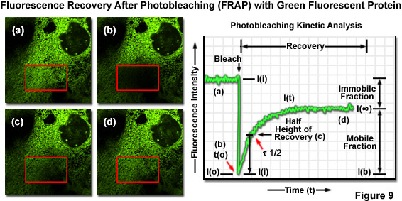
FRAP
FRAP: Fluorescence Recovery After Photobleaching
 |
|---|
| © zeiss-campus |
- Label of lipids
- Make a small dark spot with laser
- Measure the time the dark became fluorescence again.
Fluorescence recovery is due to the near lipids diffusion.
So, we can make a graph to show how long it takes to determine the diffusion-coefficient.
$$
s = \sqrt{ 4Dt}
$$
$s$: Average distance traveled
$D$: Diffusion Coefficient
$t$: Time
Membrane proteins
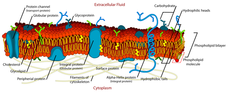 |
|---|
| © teaching.ncl.ac.uk |
Protein Diffusion
The diffusion velocity of the protein is variable.
- Similar to lipids: Photoreceptor.
- Very slow: chloride bicarbonate in red blood cell, which has interaction in the intracellular region of other protein networks, membrane skeleton.
Protein Functions
The total concentration and composition of the protein are highly variable.
Protein Position
- Integral membrane protein: fully embedded in the lipid.
- Peripheral protein: with lipids that are inserted into the lipid bilayer.
Functions
- Signal Transduction
- Molecular transportation (Pumps / Channels)
- Ion
- Glucose
- Water
Signal Role of the lipids
Both too little and too many lipids were causing pathologies.
Lipid droplets:
- Few:
Lipodystrophy & Cachexia
Lipids Maintain
The fate of lipid droplets
The form of the lipid droplets is controlled by the rate of triacylglycerol synthesis and the rate of mobilization by lipolysis
High energy state:
- GPAT, AGPAT, and MGAT catalyze the addition of fatty acids to glycerol or to monoacylglycerol to produce diacylglycerol
- DGAT1 and DGAT2 contributed to the final synthesis of triacylglycerol
- triacylglycerol stored in the endoplasmic reticulum
- triacylglycerol will then accumulate between the two leaflets of the ER to form a small bump
- Finally buds off and became a lipid droplet.
Genetic mutation reducing the activity of the AGPAT is implicated in over 50% of congenital generalized lipodystrophy.
Low energy state:
Any mutation leading to uncontrolled activation of lipolysis will also result in lipodystrophy.
Exp: mutations prevent perilipin inhibition of ABHD5 binding to the lipase ATGL, leading to constitutive activation of lipolysis, and ultimately to lipodystrophy.
Acquired Lipodystrophy
Exp: Autoimmune reaction and drug treatment.
HIV-associated acquired lipodystrophy.
Nucleoside reverse transcriptase inhibitors: mitochondrial toxicity.
Protease inhibitors: inhibit a protein needed for the processing of the nuclear protein lamin A.
High Body Fat Situation
Two diseases: Neutral Lipid Storage Disease; Metabolic Syndrome
Neutral Lipid Storage Disease
Adipocytes: Very large lipid droplets; Dominated by the TAG.
Monogenic Disease
Two Types of NLSD:
- Cardiomyopathy: Characterized by a type of heart disorder
- NLSD-M: Characterized by Ichthyosis (Sin takes on fish-scale-like appearance)
- NLSD-I
NLSD-M
The associated gene is ATGL, which catalyzes the first step of triacylglycerol.
Mutation of this gene prevented its recruitment.
Connection with Cardiomyopathy:
This mutation impairment the release of energy fuels and impacts the tissues such as the cardiac muscle.
NLSDI
NLSD-I: Chanarin-Dorfman Syndrome
Associated gene: $\alpha/\beta$-hydrolase domain-containing protein 5 (ABHD5)
ABHD5: triacylglycerol to diacylglycerol
Ichthyosis:
- “Ichthyo”: Having to do with fish
- “-osis”: pathology, or abnormal.
- The skin of the patient was dry and rough like fish scales
Causes:
- Abundant abnormal lipid deposits in the skin, which could reduce the permeability of the skin.
- The outer layer of the skin was made by lipid-based Extracellular Matrix
- The permeability of this layer is functional when lipolysis is functional.
- Impaired lipolysis leads abnormal deposition of triacylglycerol.
- Result: breakdown of the mortar holding the bricks; makes the skin taking on a dry, rough, and scale-like appearance.
Metabolix Syndrome
Metabolix Syndrome is characterized by a cluster of pathologies: high blood level of triacylglycerol, cholesterol, and LDL, hyperglycemia, and insulin resistance.
Polygenic disease.
Ectopic fat deposition -[Lipotoxity; inflammation]-> insulin resistance -> Type 2 diabetes
perilipin: control the size of the lipid droplets. (by initiating a chain reaction to lipid degradation.)
inflammation:
Principles of Biochemistry 10 |Lipids Related Membrane-Function and Pathology| Class Notes |HarvardX
https://karobben.github.io/2021/04/14/LearnNotes/edx-biochm-10/


