
Protein Folding|Graduate Biochemistry 5| Tulane
Protein Stability and Folding
Overview
- Post translational modifications
- The interactions that drive protein folding
- The interactions that drive protein unfolding
- The net stability of folded proteins
- Free energies of protein folding
- Mutation studies
- The Levinthal paradox
- Folding pathways
- Molecular Chaperones
- Degradation of proteins
Reading: All of Chapter 6
Proteolysis
- segments-cleave
- signal peptide cleaved in Membrane/secreted proteins
- proproteins: hormones; enzyems
- The final hormone is cleaved
- Some proteases are synthesized as zymogens which are activated by cleavage of a segment from the protein
Significant: Table (half life), soluable
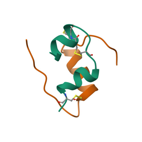
Exp: Insulin
PS: Some maturation of proteins are involved with pro-peptides cleavage. Insulin is an classical example.
Insulin has not much of structure amino acid, lack of an hydrophobic core.
6 Cystines and form 3 pair of disulfide bonds. Insulin’s primary structure has not enough information to drive it to the native structure. As a result, people believe that insulin has a memory of each processes to fold.
 |
|---|
| © PDB ID=2hiu |
Amino Acid Modulation
| Modulation Type | Added Group | Location / targets | Reversible / Not | General Features |
|---|---|---|---|---|
| Glycosylation | carbohydrates | |||
| Hydroxylation | Carboxyl | Irreversible | ||
| Methylation | Methyl | Reversible | ||
| Acetylation/Acylation | R-Carboxyl (myristate) | |||
| Carboxylation | Carboxyl | |||
| Sulfation | Sulfate | Permenent | ||
| Prenylation | Isoprenyl chains | |||
| Amidation | Amnie group | |||
| Phosphorylation | Phosphate groups | Reversible |
 |
 |
|---|---|
| © Raimund Nagel, et al | © Wiki |
Protein Folding
Interactions
The forces that stabilize folded proteins
- The hydrophobic effect
- Hydrogen bonding
- Ionic interactions
- van der Waals interactions (London dispersion force)
- Disulfide crosslinks
cite: Kauzmann, W. (1959) “Some factors in the interpretation of protein unfolding” Adv. In Proteins Chemistry. 14:1.
The hydrophobic effect
- The unfavorable interaction between hydrophobic sidechains and water is reduced when a protein folds into its native structure
- Up to 75% of hydrophobic residues are buried in folded proteins
- Its contribution greatly favors folded state
Hydrogen bonding
- Every residue in a protein has at least one H-bond donor and at least one H-bond acceptor (the peptide bond)
- Concentration of H-bonding groups in a protein is ~25M
- Most potential H-bonding groups are, in fact, H-bonded in the interior of folded proteins
- H-bonds define secondary structure geometry
The forces that stabilize folded protein
- The hydrophobic effect
- Hydrogen bonding
- Ionic interactions
- van der Waals interactions (London dispersion force)
- Disulfide crosslinks
Ionic interactions
- Salt bridges form between acidic and basic residues
- Majority of salt bridges are on surface
- Buried salt bridges are conserved
van der Waals Interaction
- Contribution is from tight packing of protein core
- Protein density is ~1.3 g/ml (H20=1.0 g/ml Ethanol=0.8 g/ml)
Disulfide Crosslinking
- Covalent crosslinks between the sulfurs of cysteine residues
- Generally nonlocal
- Significant (but not dominant) contribution to stability
The forces that destabilize folded protein
- Conformational entropy
- Hydrogen bonds to water
- Electrostatic repulsion
Conformational Entropy
- Backbone of folded protein has only one (or a few) conformations.
- The backbone of an unfolded protein has 32n conformations (because there are 3 low energy φ and ψ torsion angles for each Cα)
The backbone entropy change favoring unfolding:
ΔS = Rln(32n/1) = 2nRln(3) = 1.4 kcal/mol/residue (5.4 kJ/mol/residue)
Hydrogen bonds to water
- Every amino acid has at least two hydrogen-bonding groups
- The concentration of bulk water is ~55 Molar and each water molecule has 4 hydrogen bonding groups. Therefore the favorable energetic contribution of hydrogen bonds to folding may be small
However, the unfavorable cost of unmade hydrogen bonds in the interior of the protein is still high
Hydrophobic amino acids are buried
| Residue | Exposed | Burued | Interm ediate |
|---|---|---|---|
| S | 0.70 | 0.20 | 0.10 |
| T | 0.71 | 0.16 | 0.13 |
| A | 0.48 | 0.35 | 0.17 |
| G | 0.51 | 0.36 | 0.13 |
| P | 0.78 | 0.13 | 0.09 |
| C | 0.32 | 0.54 | 0.14 |
| D | 0.81 | 0.09 | 0.10 |
| E | 0.93 | 0.04 | 0.03 |
| Q | 0.81 | 0.10 | 0.09 |
| N | 0.82 | 0.10 | 0.08 |
| L | 0.41 | 0.49 | 0.10 |
| I | 0.39 | 0.47 | 0.14 |
| V | 0.40 | 0.50 | 0.10 |
| M | 0.44 | 0.20 | 0.36 |
| F | 0.42 | 0.42 | 0.16 |
| Y | 0.67 | 0.20 | 0.13 |
| W | 0.49 | 0.44 | 0.07 |
| K | 0.93 | 0.02 | 0.05 |
| R | 0.84 | 0.05 | 0.11 |
| H | 0.66 | 0.19 | 0.15 |
C: Buried Residues
V; L; I; F; W; C
0.01: Exposed Residues
D; E; N; Q; K; R
Protein Unfolding
- An unfolded protein has lost its tertiary/quaternary structure and its biological activity
- Depending on the experimental conditions an unfolded protein may be stable or it may aggregate/precipitate
Treatments that unfold proteins:
High temperature: many proteins unfold at 50-90 º C
pH: some proteins will unfold when they gain a large net charge
Denaturants: proteins unfold in detergents like sodium dodecyl sulfate (SDS) or in denaturants like urea and guanidine
Unfolding curves are sigmoidal
 |
|---|
| © Agnishwar Girigoswami |
Net Stability
- Most proteins have a net stability of ~10 kcal/mol/protein
Compare this to the individual contributions:- Hydrophobic effect ~ 1-2 kcal/mol/amino acid residue
- Conformational Entropy ~1-2 kcal/mol/amino acid residue
For a 200 residue protein these contributions can be 400 kcal/mol each but the overall stability will always be ~10 kcal/mol
Mutational Studies of Protein Stability
 |
|---|
| © Lydia Chang |
- Most mutations are destabilizing
- The ability to precisely predict the change in folding energy (ΔG) is not very good
- Buried residues have a bigger effect than exposed residues
[图] Predicting changes in the stability of proteins and protein complexes: a study of more than 1000 mutations. J Mol Biol. 2002 Jul 5;320(2):369-87.
The Levinthal Paradox
Does a protein find its native configuration by a random search?
Carbon-carbon bonds reorient each ~ 10-13 sec
For a protein of n residues there are 32n
conformations
If n = 100, then 32n ~ 1095
If 1013 conformations were sampled every second it would take 1082 seconds to sample them all…
…but the age of the universe is only about 1017 sec!
It is now known that proteins fold on time scales from milliseconds to minutes
- Proteins fold via conserved, hierarchical pathways
(This may explain the prevalence of the Greek Key motif)
PS:
why 32n:
- 2: there are two tortion angles: φ and ψ
- n: n peptides
- 3: If each of these bond angles can be in one of three stable conformations[1]
Diseases of protein misfolding
Cystic Fibrosis: Alzheimer’s disease
Mad Cow Disease: Retinitus pigmentosa
Molecular Chaperones
 |
|---|
| © Alexandra Richardson, 1998 |
“Heat shock proteins”
Molecular Chaperones
Chaperonins
Chaperones assist protein folding in the cell
- They bind unfolded chains
- They use energy from ATP to bind and release protein chains in a repeating cycle
- ATP turnover rate is slow 1 sec-1
The GroEL/GroES chaperone form E. Coli is well understood
Other Proteins
Protein Disulfide Isomerase
Peptidyl proline cis-trans isomerase
Protein Degradation in the Cell
There are two main pathways for protein degradation in eukaryotic cells
Lysosomal Degradation
- The lysosome is a membrane-encapsulated organelle
- It contains ~50 hydrolytic enzymes
- Lysosomes have acidic pH
- Has both selective and non-selective degradation processes
Proteasomal Degradation- The proteasome is a large protein machine (MW = 2x10⁶)
- It degrades proteins that have the small protein ubiquitin attached to them
- Ubiquitination rate (degradation rate) is determined by the N-terminal residue
- Ubiquitin (76 residues) is the most highly conserved protein known
Protein Folding|Graduate Biochemistry 5| Tulane
https://karobben.github.io/2021/09/22/LearnNotes/tulane-biochem-5/


